RESPIRATORY SYSTEM
Morphology of the upper airways

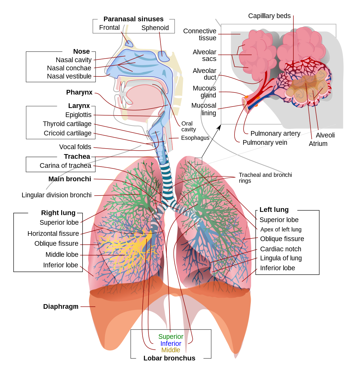
Morphology of the lower airways
*The trachea is the largest tube in the respiratory tract and consists of tracheal rings of hyaline cartilage. It branches off into two bronchial tubes, a left and a right main bronchus. The bronchi branch off into smaller sections inside the lungs, called bronchioles. These bronchioles give rise to the air sacs in the lungs called the alveoli.

*The lungs are the largest organs in the lower respiratory tract. The lungs are suspended within the pleural cavity of the thorax. The pleurae are two thin membranes, one cell layer thick, which surround the lungs. Your lungs are in your chest, and they are so large that they take up most of the space in there. You have two lungs, but they aren't the same size the way your eyes or nostrils are. Instead, the lung on the left side of your body is a bit smaller than the lung on the right. This extra space on the left leaves room for your heart.

Your lungs are protected by your rib cage, which is made up of 12 sets of ribs. These ribs are connected to your spine in your back and go around your lungs to keep them safe. Beneath the lungs is the diaphragm, a dome-shaped muscle that works with your lungs to allow you to inhale (breathe in) and exhale (breathe out) air.
From the outside, lungs are pink and a bit squishy, like a sponge. At the bottom of the trachea there are two large tubes. These tubes are called the main stem bronchio, and one heads left into the left lung, while the other heads right into the right lung.
Each main stem bronchus— the name for just one of the bronchi — then branches off into tubes, or bronchi, that get smaller and even smaller still, like branches on a big tree. The tiniest tubes are called bronchioles. Each bronchiole is about the same thickness as a hair.
At the end of each bronchiole is a special area that leads into clumps of teeny tiny air sacs called alveoli. There are about 600 million alveoli in your lungs and if you stretched them out, they would cover an entire tennis court. Now that's a load of alveoli! Each alveolus has a mesh-like covering of very small blood vessels called capillaries.

ADDED
***The alveoli are tiny air sacs in the lungs where gas exchange takes place.When the diaphragm contracts, a negative pressure is generated in the thorax and air rushes in to fill the cavity. When that happens, these sacs fill with air, making the lung expand. The alveoli are rich with capillaries, called alveolor capillaries. Here the red blood cells absorb oxygen from the air and then carry it back in the form of oxyhaemaglobin, to nourish the cells. The red blood cells also carry carbon dioxide (CO2) away from the cells in the form of carboxyhemoglobin and releases it into the alveoli through the alveolor capillaries. When the diaphragm relaxes, a positive pressure is generated in the thorax and air rushes out of the alveoli expelling the carbon dioxide.***
The gas-exchange region
The gas exchange surface of a mammal is the alveolus.There are numerous alveoli connected to the outside by the mouth and nose.These alveoli provide a massive surface area through which gases can diffuse.
These gases diffuse a very short distance between the alveolus and the blood because the lining of the lung and the capillary are both only one cell thick. The blood supply is extensive, which means that oxygen is carried away to the cells as soon as it has diffused into the blood. Ventilation movements also maintain the concentration gradients because air is regularly moving in and out of the lungs.
The upper airway consists of the pharynx and the nasal cavities; however, some authors include the larynx and trachea as well.
*The pharynx is can be divided into the nasopharynx, oropharynx, and laryngopharynx. The pharinx is a U-shaped fibromuscular tube that extends from the base of the skull to the cricoid cartilage. It is bounded anteriorly and superiorly by the nasal cavity, followed more inferiorly by the mouth, and then the larynx. These borders divide the pharynx into the nasopharynx, oropharynx, and laryngopharynx, respectively.
The epiglottis guards the opening to the glottis or the glottic inlet. It is a flap of elastic cartilage covered by mucosa that is attached superiorly and anteriorly to the larynx. Beyond the glottic inlet is the larynx.
*The nose is composed of bone and cartilage, which are in turn attached to the facial skeleton. It is a pyramidal structure that is divided by a midline septum into two nasal cavities. The nasal cavities are lined with mucosa that can function to heat and humidify inspired gas. The paranasal sinuses drain into the nasal cavity. The posterior portion of the mouth opens into the oropharynx. When a patient is supine and unconscious, the tongue and lower jaw may slide posteriorly leading to airway obstruction within the oropharynx.
* The larynx is bounded by the aryepiglottic folds, the tip of the epiglottis, and the posterior commissure of the lower border of the cricoid cartilage. It bulges posteriorly into the laryngopharynx. Beyond the cricoid cartilage lies the trachea, which is formed by a set of U-shaped cartilaginous rings that extend to the carina before bifurcating into each mainstem bronchi.
Morphology of the lower airways
*The trachea is the largest tube in the respiratory tract and consists of tracheal rings of hyaline cartilage. It branches off into two bronchial tubes, a left and a right main bronchus. The bronchi branch off into smaller sections inside the lungs, called bronchioles. These bronchioles give rise to the air sacs in the lungs called the alveoli.
*The lungs are the largest organs in the lower respiratory tract. The lungs are suspended within the pleural cavity of the thorax. The pleurae are two thin membranes, one cell layer thick, which surround the lungs. Your lungs are in your chest, and they are so large that they take up most of the space in there. You have two lungs, but they aren't the same size the way your eyes or nostrils are. Instead, the lung on the left side of your body is a bit smaller than the lung on the right. This extra space on the left leaves room for your heart.

Your lungs are protected by your rib cage, which is made up of 12 sets of ribs. These ribs are connected to your spine in your back and go around your lungs to keep them safe. Beneath the lungs is the diaphragm, a dome-shaped muscle that works with your lungs to allow you to inhale (breathe in) and exhale (breathe out) air.
From the outside, lungs are pink and a bit squishy, like a sponge. At the bottom of the trachea there are two large tubes. These tubes are called the main stem bronchio, and one heads left into the left lung, while the other heads right into the right lung.
Each main stem bronchus— the name for just one of the bronchi — then branches off into tubes, or bronchi, that get smaller and even smaller still, like branches on a big tree. The tiniest tubes are called bronchioles. Each bronchiole is about the same thickness as a hair.
At the end of each bronchiole is a special area that leads into clumps of teeny tiny air sacs called alveoli. There are about 600 million alveoli in your lungs and if you stretched them out, they would cover an entire tennis court. Now that's a load of alveoli! Each alveolus has a mesh-like covering of very small blood vessels called capillaries.
ADDED
***The alveoli are tiny air sacs in the lungs where gas exchange takes place.When the diaphragm contracts, a negative pressure is generated in the thorax and air rushes in to fill the cavity. When that happens, these sacs fill with air, making the lung expand. The alveoli are rich with capillaries, called alveolor capillaries. Here the red blood cells absorb oxygen from the air and then carry it back in the form of oxyhaemaglobin, to nourish the cells. The red blood cells also carry carbon dioxide (CO2) away from the cells in the form of carboxyhemoglobin and releases it into the alveoli through the alveolor capillaries. When the diaphragm relaxes, a positive pressure is generated in the thorax and air rushes out of the alveoli expelling the carbon dioxide.***
The gas-exchange region
The gas exchange surface of a mammal is the alveolus.There are numerous alveoli connected to the outside by the mouth and nose.These alveoli provide a massive surface area through which gases can diffuse.
These gases diffuse a very short distance between the alveolus and the blood because the lining of the lung and the capillary are both only one cell thick. The blood supply is extensive, which means that oxygen is carried away to the cells as soon as it has diffused into the blood. Ventilation movements also maintain the concentration gradients because air is regularly moving in and out of the lungs.
This breathing in (inspiration) and breathing out (expiration) is controlled via nervous impulses from the respiratory centre in the medulla of the brain.
Both the intercostal muscles (in between the ribs) and the diaphragm receive impulses from the respiratory centre. Stretch receptors in the lungs send impulses to the respiratory centre in the brain giving information about the state of the lungs.

- external intercostal muscles contract
- ribs and sternum move up and out
- width of thorax increases front to back and side to side
- diaphragm contracts
- diaphragm moves down, flattening
- depth of thorax increases top to bottom so the...
- volume of thorax increases.
- pressure between the pleural surfaces decreases.
- lungs expand to fill thoracic cavity.
- air pressure in alveoli is less than atmospheric pressure.
- air is forced in by the higher external atmospheric pressure.
As the lungs fill with air the stretch receptors send impulses to the expiratory part of the respiration centre to end breathing in.
- External intercostal muscles relax
- ribs and sternum move down and in
- width of thorax decreases front to back and side to side
- diaphragm relaxes
- diaphragm moves up
- depth of thorax decreases top to bottom.


Comentarios
Publicar un comentario