Human embryonic development
Human embryonic development
Human
development begins after the union of male and female gametes or germ cells
during a process known as fertilization. Fertilization is a
sequence of events that begins with the contact of a sperm (spermatozoon)
with a secondary oocyte (ovum) and ends with the fusion of
their pronuclei (the haploid nuclei of the sperm and ovum) and
the mingling of their chromosomes to form a new cell. This fertilized ovum, known
as a zygote, is a large diploid cell that is the beginning of
a human being
The fertilization take place in one of the Fallopian tubes. From this place the new living being, the zygote, makes a trip that finishes in the mother uterus. The uterus offers the necessary conditions for the development during the period named pregnancy.
The pregnancy is the period that spreads from the fertilization of the ovum because of the sperm to the moment of the childbirth. In the human species the period lasts around 38 and 40 weeks. During this time, the new human being passes for some changes that happen in three phases:
*Segmentation: consists of a successive cellular
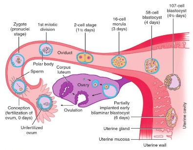 divisions for mitosis of the zygote. After
fertilization, a rapid cell division occurs
in the zygote by mitosis.
Small, spherical cells are formed from the zygote during this process, which is known as cleavage.
Small, spherical cells are formed from the zygote during this process, which is known as cleavage.
These cells are called blastomeres. The
morula develops into the blastula in the
process known as blastulation. Blastula
later becomes the embryo.
The main difference between morula and blastula is that morula is a spherical mass of
The main difference between morula and blastula is that morula is a spherical mass of
blastomeres, which are formed following
the splitting of a zygote whereas blastula
is an early developmental stage of the
embryo, consisting of a spherical layer
of cells filled with fluid.

*Morphogenesis: A continued blastula development eventually results in the gastrula. The process of conversion of a blastula into a gastrula is known as ‘gastrulation’, which is then proceeded by organogenesis.
The gastrula is comprised of three germ layers, each of which ultimately give rise to organs in the late embryo.
The three germ layers are the ectoderm, mesoderm and the endoderm. The outermost layer is the ectoderm, which later differentiates into the brain, spinal cord, skin and the nerves of the embryo. The middle layer, the mesoderm, forms connective tissues, muscles, cartilage, reproductive organs, bones, the skin dermis and teeth dentine. The innermost layer, the endoderm, differentiates into the basic primitive gut.
The three germ layers are the ectoderm, mesoderm and the endoderm. The outermost layer is the ectoderm, which later differentiates into the brain, spinal cord, skin and the nerves of the embryo. The middle layer, the mesoderm, forms connective tissues, muscles, cartilage, reproductive organs, bones, the skin dermis and teeth dentine. The innermost layer, the endoderm, differentiates into the basic primitive gut.
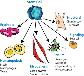
As the embryo continues to develop, individual
cells continue to differentiate. These differentiated
cell types are made from what were initially the
same types of pluripotent embryonic stem cells.
An assortment of physiological mechanisms guides
certain cells towards particular developmental
pathways, creating varying cell types.
 The embryo has 4 extraembryonic membranes:
*The amnion protects the embryo in
a sac filled with amniotic fluid.
*The yolk sac contains yolk —
the sole source of food until hatching.
Yolk is a mixture of proteins and lipoproteins.
*The chorion participates in the exchange of
O2 and CO2 between the embryo and the
outside air.
*The allantois stores metabolic wastes of the
embryo and, as it grows larger, also
participates in gas exchange.
cells continue to differentiate. These differentiated
cell types are made from what were initially the
same types of pluripotent embryonic stem cells.
An assortment of physiological mechanisms guides
certain cells towards particular developmental
pathways, creating varying cell types.
The embryo has 4 extraembryonic membranes:*The amnion protects the embryo in
a sac filled with amniotic fluid.
*The yolk sac contains yolk —
the sole source of food until hatching.
Yolk is a mixture of proteins and lipoproteins.
*The chorion participates in the exchange of
O2 and CO2 between the embryo and the
outside air.
*The allantois stores metabolic wastes of the
embryo and, as it grows larger, also
participates in gas exchange.
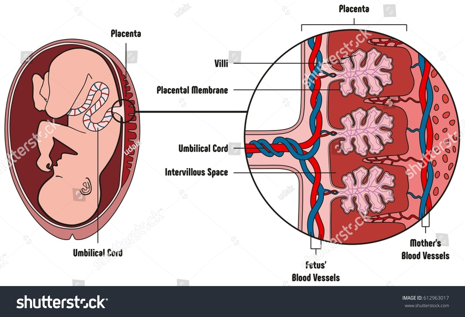
The placenta is an organ attached to the lining of the womb during pregnancy.
What does the placenta do?
Oxygen and nutrients pass from the mom’s blood supply into the placenta. From there, the umbilical cord carries the oxygen and nutrients to the unborn baby. Waste products from the baby, such as carbon dioxide, pass back along the umbilical cord to the placenta and then into your bloodstream, for your body to dispose of them.
The placenta produces hormones that help the baby grow and develop. The placenta also gives some protection against infection to the baby while it's in the womb, protecting it against most bacteria.


Comentarios
Publicar un comentario